已关闭
已关闭
- 顶部Banner位
 固定模块
固定模块

 收藏
收藏 易享客服
易享客服
The EVOS M7000 Imaging System is a high-performance, fully automated, inverted, multi-channel fluorescence and transmitted light imaging system. It was developed to meet today’s expectations for image quality, user interface interactions, data generation speed and flexibility, and end-user value. The powerful and time-saving acquisition tools with enhanced autofocus algorithms and automated routines for microwell plate assays, time-lapse live cell imaging, area scanning in time lapse and Z-stacks in scan mode, and a superb optomechanical design enable the generation of publication-quality images and data with just a few clicks when and where needed.
EVOS M7000成像系统是一款高性能、全自动、倒置、多通道荧光和透射光成像系统,其开发旨在满足当今对画质、用户交互界面、数据生成速度和灵活性以及最终用户价值的期望。强大且省时的采集工具,具有增强的自动对焦算法和用于微孔板测定、延时活细胞成像、延时扫描中的区域扫描和扫描模式下的Z-stacks的自动化例程,以及精湛的光学机械设计,只需在需要的时间和地点单击几下即可生成出版质量的图像和数据。
The EVOS M7000 Imaging System offers these advantages:EVOS M7000成像系统具有以下优点:
• Outstanding image quality and versatility with a 5-position objective turret, 4-color LED fluorescence and transmitted light channels, and 3.2 MP CMOS color and B/W cameras出色的画质和多功能性,配备5位物镜转塔、4色发光二极管荧光和透射光通道以及3.2 MP互补式金属氧化物半导体彩色和黑白相机
• Exceptional usability with fully automated X/Y scanning stage, autofocus, and acquisition routines卓越的可用性,具有全自动X/Y扫描台、自动对焦和采集程序
• High-speed image acquisition coupled with multi-position well scanning, and Z-stack and tile-stitch options lend power and flexibility to your data generation高速图像采集与多位置井扫描相结合,以及Z堆叠和瓷砖缝合选项为您的数据生成提供了强大的功能和灵活性
• Fully integrated time-lapse live cell imaging using the optional EVOS Onstage Incubator for precise control of temperature, gases for normoxic or hypoxic conditions, and humidity完全集成的延时活细胞成像,使用可选的EVOS舞台孵化器精确控制温度、常温或缺氧条件下的气体和湿度
• Powerful image analysis capabilities for cell segmentation and quantification with the optional Celleste Image Analysis Software使用可选的Celleste图像分析软件进行细胞分割和定量的强大图像分析功能
Fully automated imaging system 全自动成像系统
The EVOS M7000 system combines and integrates precision components with a unique modern design functionality that enables high-quality automated fluorescence imaging anywhere with unprecedented workflow efficiency. Full automation of the precision X/Y-stage movement; changing of the 4 LED fluorescent light cubes, 5-position objective turret, focus, and exposure; and switching of the dual camera make the EVOS M7000 system an exceptional platform for a variety of demanding imaging applications. Through the intuitive acquisition interface, you can program the EVOS M7000 system to run well-plate scanning, time lapse experiments, and tile-stitch/montage area scans in Z-stack and/or time-lapse modes in your vessel of choice. For live cell imaging over multiple hours or days, the optional EVOS Onstage Incubator, which functions as an environmental chamber on top of the microscope stage, is operated seamlessly from within the instrument interface. The EVOS M7000 system is compatible with all accessories for the predecessor EVOS FL Auto imaging system.EVOS M7000系统将精密组件与独特的现代设计功能相结合并集成,能够以前所未有的工作流程效率在任何地方实现高质量的自动化荧光成像。精密X/Y载物台移动的完全自动化;4个LED荧光灯立方体、5位物镜转台、对焦和曝光的更换;以及双摄像头的切换,使EVOS M7000系统成为各种苛刻成像应用的卓越平台。通过直观的采集界面,您可以对EVOS M7000系统进行编程,以在您选择的容器中以Z堆叠和/或延时模式运行井板扫描、延时实验和瓦片针迹/蒙太奇区域扫描。对于多小时或几天的活细胞成像,可选的EVOS Onbase孵化器(在显微镜载物台顶部充当环境室)可从仪器界面内无缝操作。EVOS M7000系统与前身EVOS FL自动成像系统的所有附件兼容。
Versatile and highly configurable 多功能且高度可配置
With a 5-position objective turret and the ability to simultaneous acquire images in four fluorescence channels plus transmitted light, the optical system can be easily configured to meet your needs. Choose from our broad range of high quality coverslip-corrected and long-working-distance objectives for use with plastic vessels or glass sides. Available from 1.25X to 100X magnification in achromat, fluorite, or apochromat models, together with over twenty LED light cubes, the sample imaging options are limitless. Dual monochrome and color high-sensitivity dual CMOS cameras enable seamless imaging of both fluorescent and chromogenically stained samples. Interchangeable vessel holders accommodate most vessel types and sizes, including slides, multi-well plates, culture flasks, and Petri dishes, and afford precise control and sample alignment by the automated stage. The vessel creation wizard in the user interface enables creation of profiles for custom sample vessels.凭借5位物镜转塔和在四个荧光通道加上透射光中同时获取图像的能力,光学系统可以轻松配置以满足您的需求。从我们广泛的高质量盖玻片校正和long-working-distance物镜中进行选择,用于塑料容器或玻璃侧面。在消色差、萤石或消色差模型中提供从1.25X到100X的放大倍率,以及超过二十个LED灯块,样品成像选项是无限的。双单色和彩色高灵敏度双互补式金属氧化物半导体相机可实现对荧光和生色染色样品的无缝成像。可互换的容器支架可容纳大多数容器类型和尺寸,包括载玻片、多孔板、培养瓶和培养皿,并通过自动化阶段提供精确控制和样品对齐。用户交互界面中的容器创建向导可以创建自定义样品容器的配置文件。
Powerful software 强大的软件
The EVOS M7000 software presents users with an extensive suite of tools for acquisition, visualization, and analysis. The user interface was designed with a strong emphasis on usability, including an option to view samples in a single field mode (Field View) or in a window that covers a much larger area (Area View). By using a zoom function, the Area View size can be adjusted. A user can explore a large sample area at low magnification, define regions of interest, and then seamlessly transition to the Scan function in Automate Mode and execute an area scan at higher magnification.EVOS M7000软件为用户提供了一套广泛的采集、可视化和分析工具。用户交互界面的设计非常强调可用性,包括在单个字段模式(字段视图)或覆盖更大区域的窗口(区域视图)中查看样本的选项。通过使用缩放功能,可以调整区域视图的大小。用户可以在低放大倍率下探索大样本区域,定义感兴趣的区域,然后在自动模式下无缝过渡到扫描功能,并在更高放大倍率下执行区域扫描。
The enhanced power of the EVOS M7000 system enables more demanding automated acquisition routines like area scanning in time lapse and Z-stacks in scan mode to be performed. Visualization capabilities include multi-field image viewing gallery, mosaic tiling and tile stitching options, Z-stack visualization, and a video recording and movie maker/editor for time lapse sequences. The EVOS M7000 system is fully compatible with the EVOS Onstage Incubator environmental chamber for control of temperature, gases, and humidity. With the ability generate both hypoxic and normoxic conditions, it is possible to study live cells over extended periods with highly flexible time lapse automation routines.EVOS M7000系统的强大功能可以执行更苛刻的自动采集程序,如延时区域扫描和扫描模式下的Z堆栈。可视化功能包括多场图像查看库、马赛克瓷砖和瓷砖拼接选项、Z堆栈可视化以及用于延时序列的视频录制和电影制作/编辑。EVOS M7000系统与EVOS舞台孵化器环境室完全兼容,用于控制温度、气体和湿度。由于能够产生缺氧和常氧条件,因此可以通过高度灵活的延时自动化程序长时间研究活细胞。
For users needing more powerful image analysis capabilities, images acquired on the EVOS M7000 system can be analyzed directly using the optional Celleste Image Analysis Software. Celleste software offers powerful tools for image segmentation and classification that can be applied to a range of cell analysis applications for counting and measuring intensity, area, and shape changes over time, and more.对于需要更强大图像分析功能的用户,可以使用可选的Celleste图像分析软件直接分析在EVOS M7000系统上获取的图像。Celleste软件提供了强大的图像分割和分类工具,可应用于一系列细胞分析应用程序,用于计数和测量强度、面积和形状随时间的变化等。
Smart LED illumination technology 智能LED照明技术
All EVOS fluorescence imaging systems utilize our proprietary LED light cubes. This light engine outputs remarkable intensity over a short light path and delivers incomparable fluorophore excitation. Each light cube contains a precisely matched set of optical components to optimize the position, evenness, and intensity of the light beam. Digitally controlled LED light sources allow adjustment of illumination levels to the sample and experimental conditions to minimize the risk of photo-toxicity and photo-bleaching in time-lapse studies. Hard-coated filters give sharper edges and significantly higher transmission efficiencies than traditional soft-coated filters.所有EVOS荧光成像系统都使用我们专有的LED光立方体。该光引擎在短光路上输出出色的强度,并提供无与伦比的荧光团激发。每个光立方体都包含一组精确匹配的光学组件,以优化光束的位置、均匀性和强度。数字控制的LED光源允许调整样品和实验条件的照明水平,以最大限度地降低延时研究中光毒性和光漂白的风险。硬涂层滤镜比传统的软涂层滤镜具有更锐利的边缘和更高的透射效率。
Easy to use and reliable 易于使用且可靠
The EVOS M7000 Imaging System is powerful, yet offers the simplicity of plug-and-play. Like other EVOS systems, it requires no warm-up or cool-down periods. The LED light source guarantees exceptional stability and durability, so you can turn the unit on and off whenever you need to image a sample. The environmentally safe, mercury-free LED bulbs are rated for >50,000 hours, compared to 300 hours for a typical mercury bulb and 1,500 hours for a metal halide bulb. The long lifetime and low energy consumption translate into significantly lower operating costs compared to instruments with conventional light sources. The EVOS M7000 system is powered by an external Dell XE4 computer with Intel Core i9 processor, 32 GB DDR4 RAM, 512 GB PCIe NVME SSD, and NVIDIA Quadro RTXA4000 with 8 GB discrete video graphics running Windows 10. Whether acquiring single images or setting up automated routines, the EVOS M7000 system is remarkably easy to use and run.EVOS M7000成像系统功能强大,但提供即插即用的简单性。与其他EVOS系统一样,它不需要预热或冷却期。LED光源保证了卓越的稳定性和耐用性,因此您可以在需要对样品进行成像时打开和关闭设备。环境安全、无汞的LED灯泡的额定寿命>50,000小时,而典型的汞灯泡为300小时,金属卤化物灯泡为1,500小时。与使用传统光源的仪器相比,长寿命和低能耗转化为显着降低的运营成本。EVOS M7000系统由外部戴尔XE4计算机供电,配备英特尔酷睿i9处理器、32 GB DDR4 RAM、512 GB PCIe NVME SSD和NVIDIA Quadro RTXA4000,带有运行Windows 10的8 GB离散视频图形。无论是获取单个图像还是设置自动化例程,EVOS M7000系统都非常易于使用和运行。
The EVOS imaging systems are built from the ground up to maximize performance and optimize workflow. You will be astonished at how easy it is to operate and amazed at how extraordinary your images look on-screen.EVOS成像系统是从头开始构建的,以最大限度地提高性能和优化工作流程。你会惊讶于它的操作是多么容易,也会惊讶于你的图像在屏幕上看起来是多么非凡。
Explore the entire EVOS line of imaging systems, accessories, and offers ›探索整个EVOS成像系统系列、配件和优惠
System and hardware specifications 系统和硬件规格
Optics:infinity‐corrected optical system; RMS‐threaded objectives with 45-mm parfocal distance光学:无限校正光学系统;RMS螺纹物镜,45毫米准焦距离
Imaging mode: fluorescence, brightfield, color brightfield, and phase contrast成像模式:荧光、明场、彩色明场和相位对比
Illumination: five position chamber for 4 fluorescence light cubes plus brightfield; light cubes with integrated hard coated filter set and LED light source with >50,000 hour life; broad selection of standard and specialty light cubes照明:用于4个荧光立方体和明场的五位置腔;带有集成硬涂层滤光片组和LED光源的立方体,寿命>50,000小时;标准和特殊立方体的广泛选择
Imaging methods: single color, multi-color, area scan with montage or tile-stitch, time lapse, Z-stacking, movie capture成像方法:单色、多色、蒙太奇或瓷砖针迹区域扫描、延时、Z堆叠、电影捕捉
Objective capacity: 5‐position turret目标容量:5位炮塔
Objectives (not included): wide selection of high‐quality LWD and coverslip‐corrected objectives目标(不包括):广泛选择高质量LWD和盖玻片校正目标
Condenser:60-mm long working distance condenser, 4 position turret with a clear aperture and 3 phase annuli冷凝器:60mm长工作距离冷凝器,4位转塔,带透明孔径和3相环空
Stage: motorized X/Y scanning stage; travel range 120 mm x 80 mm with sub‐micron resolution, drop‐in inserts to receive vessel holders and lock down holders to fix sample in place during long scans阶段:电动X/Y扫描阶段;行程范围120毫米x 80毫米,亚微米分辨率,用于接收容器支架和锁定支架的插入式插件,用于在长扫描期间将样品固定到位
Focus mechanism:automated focus mechanism with sub‐micron resolution对焦机构:具有亚微米分辨率的自动对焦机构
LCD display: 27-inch 4K color monitor; 3840 x 2160 pixel resolutionLCD显示屏:27英寸4K彩色显示器;3840 x 2160像素分辨率
Cameras: high-sensitivity 3.2 MP (2048 x 1536) monochrome CMOS sensor with 3.45 µm pixel resolution;high-sensitivity 3.2 MP (2048 x 1536)相机:高灵敏度3.2 MP(2048 x 1536)单色互补式金属氧化物半导体传感器,分辨率为3.45µm像素;高灵敏度3.2 MP(2048 x 1536)
仅供科研使用。不可用于诊断程序。
规格
类型细胞成像系统
产品线EVOS: EVOS:
相机320 万像素、彩色、CMOS(2个摄像头)
适用于(应用)神经生物学、免疫肿瘤学、活细胞成像、3D 细胞成像、高分辨率平铺扫描、免疫组织化学技术 (IHC)
型号M7000
光源LED
放大1.25X 至 100X 1.25X 100X
对象5 位转盘
分辨率1920 x 1080 像素
显示器LCD 液晶显示器
数量1 system 1个系统
运输条件室温
Unit Size 单位大小Each 每个
内容与储存
EVOS M7000 细胞成像系统
• 23英寸高分辨率触摸屏戴尔彩色显示器,1920 x 1080 像素分辨率
• 外置戴尔计算机,配备英特尔酷睿 i7-11700k 处理器,32 GB DDR4 RAM,512 GB PCIe NVME SSD,并配备运行 Windows® 10 的8 GB 离散视频图形的 NVIDIA Quadro RTX4000 独立显卡,设计用于触摸屏显示器和显微镜
EVOS M7000 配件套件
•EVOS M7000 快速入门指南
• 16GB USB 3.0 驱动及用户手册
• 通用电源,24V,5A
• 3根北美(B 型)电源线*
• USB 3.0 A 型接 B 型线缆
• 显示端口接显示端口线缆,6英尺
• 光立方访问工具
• EVOS 校准载玻片 (AMEP4720)
• 可用于固定两个载玻片 (AMEPVH021) 的容器支架
• 可用于固定微孔板 (AMEPVH022) 的容器支架。也可用作容器支架 AMEPVH001 至 AMEPVH018 的适配器。
• 阻挡聚光器滑块 (AMEP4688)• 适用于明场应用的漫射聚光器滑块 (AMEPDFS1)
• 适用于 4X 物镜的滑块 (AMEP4738)
• EVOS 遮光罩
• 防尘罩
• 运输限制解除指南
• 鼠标垫
• 技术支持卡
• USB 有线光电鼠标
• 键盘
* 请注意:在不使用 B 型(北美)电源插头的地区,必须单独订购三 (3) 根特定国家/地区的电源线:
AMEP4644 EVOS 电源线,A 型(北美)
AMEP4645 EVOS 电源线,G 型(英国)
AMEP4646 EVOS 电源线,C 型(欧洲)
AMEP4647 EVOS 电源线,I 型(澳大利亚)
AMEP4648 EVOS 电源线,L 型(意大利)
AMEP4649 EVOS 电源线,H 型(以色列)
AMEP4708 EVOS 电源线,D 型(印度/南非)
AMEP4782 电源线,I 型(中国,CCC 认证)
AMEP4786 电源线,J 型(瑞士)

新客户登录会员中心即赠100积分!开工季优惠,首单加赠500积分,限时500积分抽大奖!
图表

HeLa cells labeled with Alexa Fluor Plus 647 Phalloidin用Alexa Fluor Plus 647 Phalloidin标记的HeLa细胞

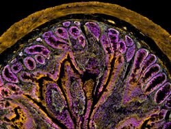
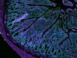
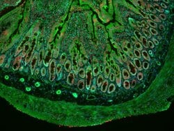
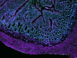
Mouse kidney labeled with Alexa Fluor Plus 555 Phalloidin用Alexa Fluor Plus 555 Phalloidin标记的小鼠肾脏
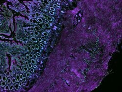
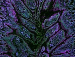
CellMask Deep Red Actin Tracking Stain labeling in IHC protocolCellMask深红色肌动蛋白跟踪染色标记在IHC协议中
CellMask Deep Red Actin Tracking Stain labeling in IHC protocolCellMask深红色肌动蛋白跟踪染色标记在IHC协议中

HeLa cells labeled with Alexa Fluor Plus 555 Phalloidin用Alexa Fluor Plus 555 Phalloidin标记的HeLa细胞

HeLa cells labeled with Alexa Fluor Plus 555 Phalloidin

HeLa cells labeled with Alexa Fluor Plus 647 Phalloidin

HeLa cells labeled with Alexa Fluor Plus 647 Phalloidin
CellMask Deep Red Actin Tracking Stain labeling in IHC protocol

HeLa cells labeled with Alexa Fluor Plus 555 Phalloidin
CellMask Deep Red Actin Tracking Stain labeling in IHC protocol
CellMask Green Actin Tracking Stain labeling in IHC protocol
CellMask Deep Red Actin Tracking Stain labeling in IHC protocol
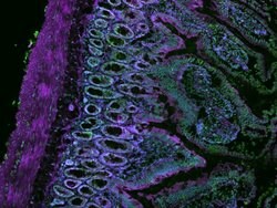
CellMask Deep Red Actin Tracking Stain labeling in IHC protocol

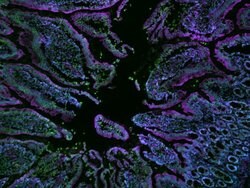
Multiplex image of live HeLa cells
CellMask Deep Red Actin Tracking Stain labeling in IHC protocol

Multiplex image live HASM cells

HeLa cells labeled with Alexa Fluor Plus 647 Phalloidin

iPSC imaged on EVOS M7000 microscope

Mouse kidney labeled with Alexa Fluor Plus 647 Phalloidin

Multiplex image of live HeLa cells

Multiplex image of ICC using HeLa cells

iPSC imaged on EVOS M7000 microscope
视频
 赛默飞mRNA疫苗整体解决方案.pdf创新药出海白皮书.pdf创新药出海白皮书.pdf化学药物分析整体解决方案.pdf化学药物分析整体解决方案.pdf化学药物分析整体解决方案.pdf赛默飞疫苗整体解决方案.pdf赛默飞制药挤出与分析解决方案.pdf中药分析完整解决方案.pdf中药分析完整解决方案.pdf赛默飞细胞与基因治疗整体解决方案.pdf赛默飞细胞与基因治疗整体解决方案.pdf赛默飞生物药解决方案概览.pdf赛默飞抗体药物整体解决方案.pdf